This website uses cookies that help the website function and that help us understand how you interact with it. Please read our privacy policy for more information.
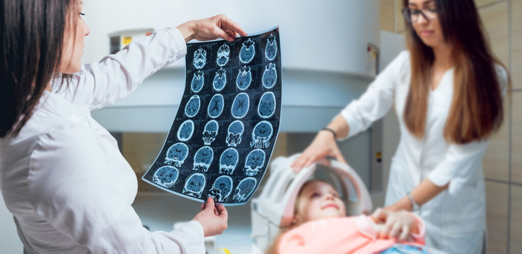
Biomarker testing is a critical step to accurately identifying a child’s brain tumor and making informed decisions about care. The process typically begins when a neurosurgeon removes tumor tissue through a biopsy or surgical resection.
Before biomarker testing begins, tumor tissue will be sent to pathology to see what the cells look like under a microscope.
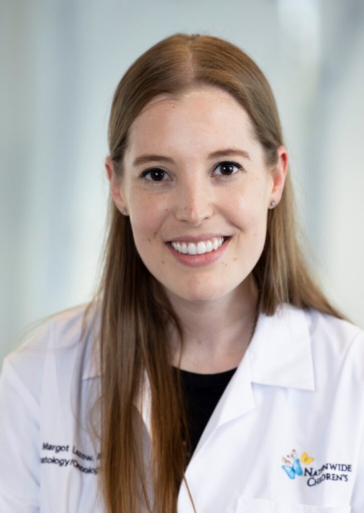
Margot Lazow, MD, MS
“Neuropathologists look at the tumor tissue under the microscope to determine the cell of origin,” said Margot Lazow, MD, MS, a pediatric neuro-oncologist at Nationwide Children’s Hospital. “For example, is it glial, neuronal, or other? Additionally, neuropathologists will assess how aggressive the tumor is, how atypical the cells are, and how rapidly tumor cells are growing or proliferating.”
The pathologist will then do special stains, called immunohistochemistry, to mark the tumor tissue for certain proteins on the cell surface or sometimes within the cell. These protein stains help guide the team’s understanding of what this tumor is and its aggressiveness.
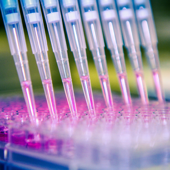
Once the pathologist has completed their analysis, the tumor tissue can then be sent off for comprehensive molecular biomarker testing.
What is Biomarker Testing?
Biomarker testing, also called molecular testing or tumor profiling, is the analysis of one’s brain tumor tissue at the molecular level to identify the genes, proteins, or other biological data that are expressed. Results from this testing can help inform the diagnosis, prognosis, and treatments available.
“Many of the biomarkers we evaluate for are different genetic alterations; gene changes that further characterize the tumor,” Dr. Lazow said. “Biomarker testing offers information about the tumor type to help confirm a specific diagnosis, while also providing insight into how aggressive the tumor is, guiding understanding of prognosis, and presenting opportunities for targeted treatment.”
Types of Biomarker Testing
The term biomarker testing represents a variety of comprehensive panels performed to understand the genetic and molecular features of the tumor. Depending on the institution, this may include single biomarker tests or broader, comprehensive panels that examine multiple biomarkers at once. Patients should feel empowered to ask their doctor which test is being used and why.
For example, the Institute for Genomic Medicine (IGM) at Nationwide Children’s Hospital offers three clinically validated molecular biomarker tests for their patients, as well as for patients around the country who are newly diagnosed with a pediatric brain tumor through the Children’s Oncology Group (COG) Project Every Child (APEC) Molecular Characterization Initiative (MCI).
1. Methylation Profiling
“Methylation evaluates the genetic profile of your child’s tumor, or more specifically, the epigenetic profile, focused on protein modifications to the DNA impacting gene expression,” Dr. Lazow said. “Through methylation testing, the epigenetic profile of your tumor is compared to databases of 10s of 1000s of other pediatric brain tumors to see which diagnosis this tumor matches the closest with and with what probability score. It provides valuable information both to confirm diagnosis and often subclassify tumors into relevant subgroups. For some pediatric brain tumors like medulloblastoma, the molecular subgroup that is determined from methylation profiling greatly informs our understanding of the disease, prognosis, and how we should treat it.”
2. A Fusion Panel
“Whereas methylation is performed using DNA extracted from the tumor, the fusion panel is performed using RNA,” Dr. Lazow shared. “This panel looks at genes that are fused — rearranged and stuck together. When you have two genes stuck together when they normally wouldn’t, you can get activation of that gene and high levels of the protein it leads to, or vice versa, depending on the location of where the genes fuse together. Certain gene fusions that we can detect not only inform our understanding of the tumor type, but also potentially can be targeted with targeted therapies. For example, low-grade gliomas often have BRAF fusions, and we can potentially target these alterations with MEK inhibitors or Pan-RAF inhibitors.”
3. Whole Exome Sequencing
This test looks at nearly all of a patient’s genes by sequencing both tumor tissue and a sample of healthy cells, usually from blood. By comparing the two, doctors can identify which genetic changes are unique to the tumor (called somatic mutations) and whether any inherited mutations (called germline mutations) might increase a child’s risk of developing cancer.
“For most brain tumors that we treat in children, there’s not an inherited cause,” Dr. Lazow said. “However, in a minority of patients, there may be a genetic alteration passed down through the family that increases tumor risk, which can be important to identify.”
For example, there are rare conditions in a group called mismatch repair deficiency, which is a type of inherited genetic predisposition that puts patients at a higher risk for cancers because the cells don’t repair their DNA appropriately.
“Brain tumors in the setting of underlying mismatch repair deficiency are more sensitive to a certain type of immunotherapy called immune checkpoint inhibitors,” Dr. Lazow said. “Therefore, in rare cases, we can take advantage of the inherited genetic predisposition for treatment decisions, which is valuable information to learn.”
In addition, discovering a genetic predisposition can inform family testing, guide long-term surveillance, and even affect treatment decisions if they have a higher risk of secondary cancers from a particular therapy.
What are Common Biomarkers of Pediatric Brain Tumors?
Common genetic and molecular features of children’s brain tumors include gene fusions like BRAF or NTRK, mutations such as BRAF V600E or H3K27M, and gene amplifications like MYC or MYCN. Identifying these changes early at diagnosis can guide personalized treatment plans and help families make informed decisions about care.
“For several brain tumors, there are certain molecular biomarkers that you can take advantage of from a therapeutic standpoint, such as targeting with oral targeted therapies,” Dr. Lazow said.
Low-Grade Gliomas
Low-grade gliomas, which are the most common brain tumor in children according to the National Cancer Institute, can have distinct biomarkers that can be matched with targeted therapies.
“Low-grade gliomas generally have one single driver genetic change,” Dr. Lazow said. “Their genome is less chaotic than the high-grade tumors, which often have several co-occurring genetic alterations.”
BRAF Fusions
A BRAF fusion happens when two genes that don’t normally connect get rearranged and stuck together like two electrical wires spliced incorrectly. This faulty wiring creates a constant growth signal in the tumor cells, telling them to keep dividing even when they should stop. According to the National Cancer Institute, “fusions are the most common changes in BRAF that occur in children and teens with low-grade gliomas.”
Researchers have developed medications that can target this gene fusion. MEK inhibitors (such as trametinib, selumetinib, or binimetinib) and a newer class of drugs called pan-RAF inhibitors (including the FDA-approved tovorafenib) can sometimes help slow or stop tumor growth when a BRAF fusion is present.
Dr. Lazow said, “If your patient has a pilocytic astrocytoma with a BRAF fusion and treatment is indicated, such as at progression or recurrence, there could be consideration of the MEK or pan-RAF inhibitor.”
However, clinical trials are underway comparing chemotherapy with these newer targeted therapies in newly diagnosed patients. The results of these studies will help doctors determine whether targeted drugs eventually become the preferred first treatment.
What does this mean for families? If a child’s tumor has a BRAF fusion, it opens the door to additional treatment possibilities beyond traditional chemotherapy. While chemotherapy remains the usual first step, targeted drugs are already available in some situations and are being studied in clinical trials. This makes biomarker testing especially important because it helps doctors identify whether these newer, potentially better-tolerated options could benefit the child, now or in the future.
BRAF Mutation
Unlike BRAF fusions, which involve two genes being stuck together, BRAF mutations are different because it’s a change within the gene itself. The most common mutation in pediatric low-grade gliomas is the BRAF V600E mutation.
This mutation acts like a car with the gas pedal stuck to the floor, constantly accelerating and pushing out growth signals. Because the tumor cells can’t slow down, they keep dividing unchecked.
“If your child has a BRAF V600E mutant low-grade glioma, we now have clinical trial data that’s been published demonstrating that oral BRAF inhibitor, in combination with MEK inhibitor, is superior to chemotherapy when studied head to head with regard to response rate and disease control,” Dr. Lazow said. “Not all low-grade gliomas need systemic therapy, but if treatment is indicated, we would now recommend the BRAF/MEK inhibitor combo (dabrafenib and trametinib) [or even consideration of oral pan-RAF inhibitor tovorafenib] over chemotherapy first-line.”
What does this mean for families? If your child’s tumor has a BRAF V600E mutation, doctors may recommend targeted therapy with a BRAF/MEK inhibitor combination or an oral pan-RAF inhibitor instead of chemotherapy. Knowing whether this mutation is present provides a clearer roadmap for treatment.
FGFR Alterations
According to a study in Nature Communications, 8.9% of pediatric gliomas have changes in the fibroblast growth factor receptor (FGFR) family of proteins. These FGFR alterations, typically in FGFR1 or FGFR2 genes, can take the form of mutations or fusions, and they act like a “stuck signal” telling cells to grow continuously, similar to a gas pedal that won’t let up, though a different part of the growth pathway is affected.
“There are targeted FGFR inhibitors, but we’re still learning as a community how effective they are for FGFR-altered low-grade and high-grade gliomas and how safe they are given some unique toxicities,” Dr. Lazow said. “We are also still learning if they are similarly effective for all FGFR alterations such as fusions, internal tandem duplications, mutations, etc. More to come as this is still under investigation for pediatric brain tumors.”
What does this mean for families? If a child’s tumor has an FGFR alteration, it may open the door to specialized treatments being tested in clinical trials. Biomarker testing at diagnosis is crucial for identifying these changes and ensuring the child has access to all available options as research advances and new discoveries are made.
High-Grade Tumors
Pediatric high-grade gliomas are a diverse and aggressive group of brain tumors. Each tumor can have different genetic changes, which means prognosis and treatment options can vary, though in general unfortunately outcomes are poor. Biomarker testing of the tumor’s genetic and molecular features is critical to identify these alterations and guide potential targeted therapies.
As Dr. Lazow explains, “High-grade gliomas represent a heterogeneous disease of aggressive glial tumors, comprised of different molecular subgroups, often with distinct genetic alterations. Molecularly-targeted therapy may be one part of a multi-step (multimodal) treatment plan for pediatric high-grade gliomas that also involves radiation as well as consideration, in some cases, of immunotherapy.”
NTRK Fusion
Although less common, some pediatric low-grade and high-grade brain tumors have a neurotrophic tyrosine receptor kinase (NTRK) fusion. This occurs when the NTRK gene is abnormally fused with another gene, resulting in a faulty signal that drives tumor growth.
An Acta Neuropathologica Communications article states, “Approximately 0.55 to 2% of all gliomas/neuroepithelial tumors contain NTRK fusions, though the incidence may be up to 5.3% in pediatric high-grade gliomas (HGG), 4% of diffuse intrinsic pontine gliomas (DIPG), and 40% of non-brainstem HGG in patients younger than 3 years old.”
Leon
Solid tumors with NTRK fusions, including brain tumors, can be targeted with medicines called TRK inhibitors, such as larotrectinib or entrectinib. These drugs have shown strong responses in children with tumors driven by NTRK fusions and are now FDA approved for this indication, offering another example of how biomarker testing results can guide treatment.
What does this mean for families? While rare, if an NTRK fusion is found in a child’s tumor, it opens the door to a highly targeted therapy option that may be more effective and less toxic than traditional treatments.
Four-month-old Leon was able to take liquid medication (larotrectinib) for his high-grade astrocytoma with NTRK fusion instead of undergoing chemotherapy.
“Thank God that there’s this drug out there that specifically targets this mutation,” said David, Leon’s dad. “It’s wild that there’s such a medication sitting in our fridge next to the celery and sour cream.”
H3K27M Mutation
The H3K27M mutation is a change in a gene that helps control how DNA is packaged and how certain genes are turned “on” or “off.” Specifically, it affects histone proteins — proteins that provide structural support for a chromosome. Normally, histones help regulate which genes are active and when. When the H3K27M mutation occurs, it disrupts this regulation. Genes that should be quiet can become inappropriately active, and this abnormal signaling drives tumor growth.
Diffuse midline glioma (DMG), H3K27-altered (which also includes DIPG when in the pons), is a rare and aggressive brain tumor of midline location (brainstem, thalamus, spinal cord) that mostly affects children and young adults. About 90% of these tumors have the H3K27M mutation, according to Neuro-Oncology.
There are sadly no curative therapies for children diagnosed with DMG. Standard treatments like radiation can temporarily slow the disease and often provide symptom benefit, but families are left with very few options once the tumor progresses. The FDA recently approved dordaviprone (Modeyso) for patients with recurrent H3K27M-mutant DMG. There are also many ongoing clinical trials of biologically-targeted therapies and immunotherapies being studied in this disease.
“We hope the community can see this approval as the beginning of a new era in treatment development for DMG, and more broadly, rare and recalcitrant cancers,” said David Arons, President & CEO for the National Brain Tumor Society. “While outcomes remain difficult for many patients, this therapy introduces a new option based on the underlying biology of the disease and patients’ biomarkers. There is much work still to be done to improve outcomes, understand which patients are most likely to benefit, and develop additional therapies.”
What does this mean for families? While this breakthrough does not represent a cure, it is the first targeted therapy to make it through regulatory approval for this devastating disease.
Research is ongoing to understand who benefits most from this drug, and the approval gives families an additional option and, importantly, a sense of hope in an area where hope has been scarce.
MYC and MYCN Amplification
MYC and MYCN are part of the MYC family of genes. Think of them as master controllers inside cells that help decide when a cell should grow, divide, or mature into its final form. While MYC works in many types of cells, MYCN (sometimes called N-Myc) is especially important during brain and nervous system development, where it helps young nerve cells grow properly.
When functioning normally, MYC and MYCN act like skilled orchestra conductors, making sure all the growth-related genes stay in sync. But sometimes tumors develop extra copies of one of these genes — a change called amplification. When this happens, it’s like dozens of extra conductors show up, all frantically waving their batons. This can overwhelm the normal checks and balances in the cell, leading to runaway growth and cancer.
Right now, there are no FDA-approved drugs that directly turn off MYC or MYCN. But researchers are exploring new therapies that could destabilize the protein or interfere with the signals it sends.
What does this mean for families? While therapies targeting MYC and MYCN are still in development, biomarker testing for these amplifications is important because it gives doctors a better understanding of how aggressive the tumor might be and can help guide enrollment in clinical trials exploring new treatments.
Gene Fusions in Infant Hemispheric Gliomas
Some rare but aggressive brain tumors that occur in very young children are called infant hemispheric gliomas. These generally high-grade tumors arise in the cerebral hemispheres — the large right and left sides of the brain.
Researchers have discovered that many of these tumors are driven by gene fusions. As described above, gene fusions happen when two different genes accidentally join together. Common fusions in infant hemispheric gliomas include NTRK, ROS1, ALK, and MET genes.
Many of these fusions are potentially targetable with existing cancer drugs called targeted therapies (for example, NTRK inhibitors or ALK inhibitors), but more research is needed to know the best way to use these drugs in children — whether right away, at recurrence, or in combination with chemotherapy.
“We are still studying the ideal sequence and combination of therapies for infant hemispheric gliomas, recognizing they often have a clear molecular target through these gene fusions for which targeted therapy can be considered at some point in their disease course,” Dr. Lazow said. “We’re still learning the best way to optimize, sequence, and potentially even combine targeted therapies with conventional chemotherapy, including through ongoing clinical trials.”
What does this mean for families? For families whose child is diagnosed with an infant hemispheric glioma, biomarker testing is important to know whether a targetable fusion is present. If the tumor has one of these gene fusions (such as NTRK, ROS1, ALK, or MET), there may be the possibility of using a targeted therapy.
Can Biomarker Testing Impact Clinical Trial Opportunities?
Biomarker testing doesn’t just help guide current treatment — it can also determine eligibility for clinical trials. Many pediatric brain tumor trials now require or prioritize patients whose tumors have specific genetic changes.
“Some clinical trials, especially of molecularly-guided therapy, rely on the presence or absence of a genetic biomarker, so this may impact trial eligibility,” Dr. Lazow said.
Knowing your child’s biomarkers early means doctors can identify trials that are a good fit and that may offer access to promising new therapies that wouldn’t otherwise be available. This is especially important for high-grade tumors or rare subtypes, where standard options are limited and targeted treatments could make a difference.
Precision Medicine for High-Grade Gliomas
NBTS has provided several years of funding, expertise, and advice to support the CONNECT Consortium, an international collaborative network of pediatric cancer centers focused on improving outcomes for children and young adults with high-risk brain tumors, including high-grade gliomas, diffuse intrinsic pontine glioma, medulloblastoma, ATRT, and craniopharyngioma.
Their new molecularly-guided phase II umbrella trial, CONNECT TarGeT, offers an innovative precision medicine approach for children and young adults newly diagnosed with high-grade gliomas, including DIPG and other DMGs. All participants undergo comprehensive molecular testing of their tumor. Patients are then assigned to one of several targeted therapy arms, often used alongside or after upfront radiation.
Dr. Lazow explains, “All patients undergo detailed molecular testing of their tumor upfront, and then patients are assigned to targeted therapy arms based on and matching to the unique genetic profile of their tumor. Efficacy of these targeted treatments is being studied in this phase 2 umbrella trial.”
Join NBTS for a Free Biomarker Testing Webinar on Sept. 26, 2025
When you ID your tumor through biomarker testing, you can explore treatment options with your health care team and make informed decisions about your care, including whether a clinical trial may be right for you. With MyTumorID, I Decide.
Join us for a free informative MyTumorID webinar on Sept. 26, 2025, featuring an expert in the field who will explain what biomarker testing is, how it influences treatment planning and access to clinical trials, and why it matters. You’ll also hear from a patient about how biomarker testing results impacted their brain tumor experience. This educational webinar will conclude with a moderated discussion of questions submitted by the audience.