This website uses cookies that help the website function and that help us understand how you interact with it. Please read our privacy policy for more information.
News stories about patients playing instruments, such as a saxophone or guitar, or singing Taylor Swift songs during brain surgery often make headlines and go viral on social media. These dramatic moments typically occur during a highly specialized procedure known as an awake craniotomy.
A craniotomy is a surgical procedure in which a bone flap is removed from the skull to access the brain for a variety of brain-related conditions, including brain tumor resection, when part or all of the tumor is removed. Depending on the brain tumor’s location, a neurosurgeon may determine that an awake craniotomy is needed rather than the traditional sedated craniotomy, where the patient is fully unconscious under general anesthesia.
For individuals preparing for this kind of procedure, the viral videos only scratch the surface. What does it mean to be awake during brain surgery, and why do surgeons choose this method in the first place?
This guide walks through what patients can expect before and during an awake craniotomy.
Why Do Neurosurgeons Perform an Awake Craniotomy?
The primary goal of a brain tumor resection is to remove as much of the tumor as possible while preserving essential brain functions through individualized mapping techniques. When a tumor is located near areas of the brain that control speech, language, movement, or vision, an awake craniotomy allows the surgical team to monitor and protect those functions in real time.
By keeping the patient awake and engaged during specific parts of the operation, the neurosurgeon can map key functions in the brain and test critical areas before making surgical changes.
“An awake craniotomy gives the surgeon really good feedback in the moment instead of just a before and after picture,” said Nate Somes, MS, CCC-SLP, of the Massachusetts General Hospital Speech Language Pathology Brain Tumor Team. Speech language pathologists (SLPs) may be present in the operating room or prep patients before awake craniotomies if a tumor is located in the speech and language area of the brain.
Keep in mind that an awake craniotomy isn’t always necessary or the “better” technique across the board. This technique is only needed when preserving brain function requires direct communication between the patient and the surgical team during the operation.
Aaron Cohen-Gadol, MD
“I have a philosophy that an awake craniotomy is a big deal for the patient; therefore, it should only be done if it’s absolutely necessary,” said neurosurgeon Aaron Cohen-Gadol, MD. “If the tumor is very much infiltrating the speech area of the brain or the motor or sensory cortex or cortices, those are the best candidates. I’ve learned that even when the tumor comes close, but it’s not infiltrating, I have been able to remove the tumor effectively without an awake craniotomy, and it’s much easier on the patient.”
Gliomas tend to be the most common tumor type to warrant an awake craniotomy. According to theJournal of Neurosurgery, “an estimated 46-61% of gliomas are located within presumed eloquent regions of the brain” — areas believed to control critical functions like speech, movement, or memory.
Surgeons will also consider the patient’s overall health and whether the patient is willing to be awake during surgery and cooperate with the questions and tasks being asked of them.
“Not every patient is a candidate for an awake craniotomy and not every tumor location is in the right spot to even consider it,” said Amy Maguire, PhD, CCC-SLP, of the Massachusetts General Hospital Speech Language Pathology Brain Tumor Team. “Individual surgeons often have varying perspectives and approaches to doing an awake surgery, so they might have different criteria for recommending it [to a patient]. When our SLP team is involved in awake surgeries, we spend a substantial amount of time with patients before surgery to help them feel well-prepared, in addition to completing a pre-surgical evaluation. We try to make ourselves as available as possible in the lead-up to surgery, knowing that patients and families often have questions that pop up after their scheduled visits.”
Seeking a Second Opinion
An awake craniotomy is a specialized procedure, so it should be done by a neurosurgeon who has significant experience with this particular technique. Smaller institutions may perform only a handful of awake craniotomies each year, whereas some NCI-Designated Cancer Centers perform nearly 100 awake craniotomies annually. Unless the patient is experiencing a true emergency, there is time to seek a second opinion to ensure an awake craniotomy is the best course of action and that the surgery will be done by someone with the necessary experience.
“I think second opinions are very important,” Dr. Cohen-Gadol (Cohen) said. “Most patients who are diagnosed end up seeing a family doctor. They’re referred to a neurosurgeon, and they’re not able to recognize which neurosurgeon is actually the best one to perform the operation. I highly recommend researching your surgeon online. Know how many of these surgeries they do and if they have specialized training in the removal of these tumors, because the outcomes are definitely better, especially for more difficult tumors. Your health is your most important asset. Therefore, you should get the best surgeon to take care of you.”
What to Expect Before the Awake Craniotomy
The process leading up to an awake craniotomy involves careful preparation and a team approach to ensure the patient is physically and emotionally ready. Every surgical team has slightly different workflows, but most patients will go through two key steps before the procedure, including a pre-operative appointment and baseline testing for brain mapping.
Pre-op Discussion With Your Neurosurgeon
Before surgery, patients typically meet with their neurosurgeon to walk through the procedure and address any concerns. This appointment provides an opportunity to clarify what will happen during surgery and how the team will work to keep the patient safe and comfortable.
“We try to have a session where we talk the patient through the process before surgery,” Dr. Cohen said. “The more the patient knows, the more comfortable they are, the more they can expect. This detailed session will tell them what we’re doing, what could bother them, and why they should reach out to us if anything is bothering them.”
Topics frequently discussed during this visit include:
What the operating room will look and feel like
How and when the patient will be awake during the procedure
Specific tasks or language exercises the patient may be asked to do during surgery
Pain management concerns
Whether the patient has claustrophobia or other concerns
Potential risks and benefits of the procedure
What abilities and skills are important to the patient’s life to help inform the neurosurgeon on their surgical approach, and when resection should be stopped
Recovery expectations
“My husband didn’t want a catheter,” said Stephanie* about a concern that her husband brought up in his pre-op appointment. “His surgeon worked with him, and they agreed to put a funnel there, and he just had to tell them when he had to pee so they could help. I loved that level of care.”
While avoiding a catheter may not be possible, it’s an example of the type of concerns a patient can bring up to their team for discussion ahead of surgery.
Patients are also encouraged to ask their own questions during this pre-op appointment. Some of the most common questions about awake craniotomies include:
Why do I need to be awake during brain surgery?
Will I feel pain during surgery?
Will I be fully awake and aware the whole time?
What will you need me to do when I’m awake?
What happens if I panic or get claustrophobic?
How will my head be kept still?
What are the risks and side effects?
What should I expect during recovery?
“Looking back, the hardest part was the night before [surgery] — all the things I envisioned,” shared Brian S., who had an awake craniotomy for an astrocytoma resection. “I was worried that if I’m going to be awake, and they’re in my head, I’m going to feel pain or them sticking something in my brain. Just the unknown is scary.”
By discussing concerns and asking questions ahead of the surgery, patients can help minimize their pre-op concerns.
Functional Mapping Preparation
An essential part of the awake craniotomy is awake brain mapping, a technique used to identify and preserve critical brain functions. Before undergoing surgery, the surgical team requires an understanding of the patient’s baseline cognitive and language abilities.
Patients often work with speech-language pathologists (SLPs) or neurologists before surgery to do this baseline testing. This session may involve flashcards, picture naming, reading tasks, answering questions, or movement exercises — most of which will be used again during surgery.
“We help patients get familiar with the language tasks they’ll do during surgery,” said Sarah Steele, MS, CCC-SLP, of the Massachusetts General Hospital Speech Language Pathology Brain Tumor Team. “The goal is to make sure there are really no surprises and to help them gain some familiarity with the types of questions, pictures, or reading tasks that they’ll see.”
Familiarity with the test materials is important — not just for the patient to feel comfortable going into surgery, but to help the surgical team accurately interpret any changes in the patient’s responses during the operation.
Language Area Mapping
For tumors near critical language areas, the baseline testing will focus on tasks that can help determine if the stimulation during brain mapping interferes with the ability to speak clearly, find words, or understand language. A 2021 article in Neurosurgery included a figure that outlines the preoperative and intraoperative cognitive tests frequently used depending on the location of the tumor.
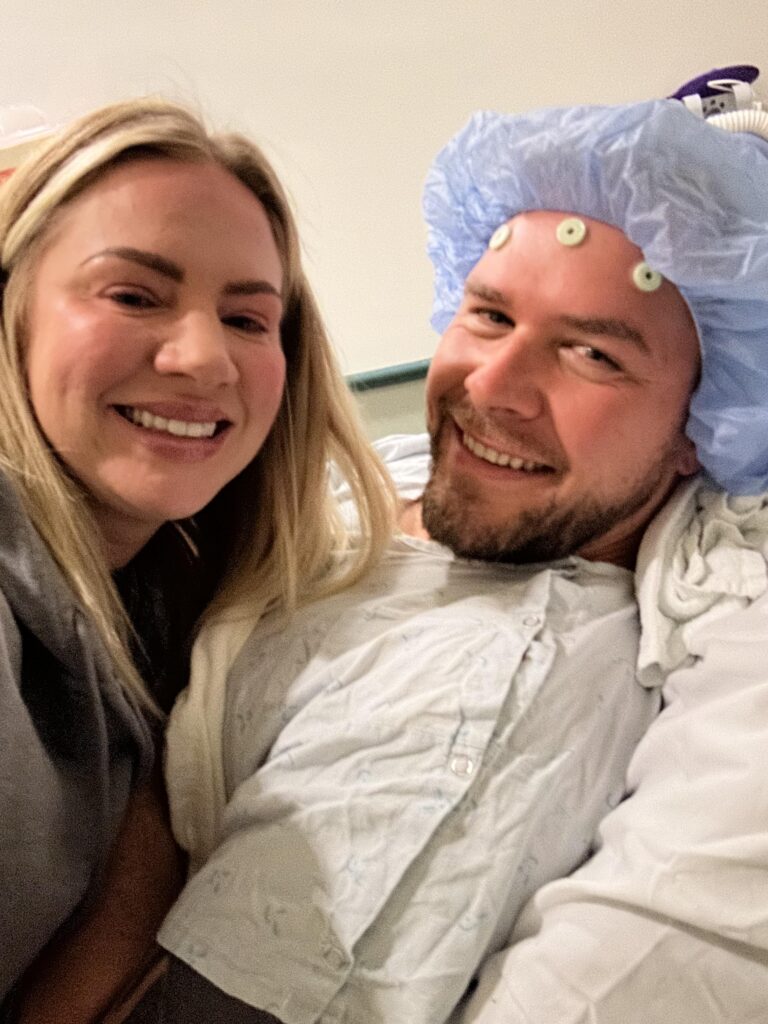
“We had to meet with the neurologist who was going to be doing the brain mapping,” said Katie N., whose husband Mike had an awake craniotomy for oligodendroglioma. “The doctor asked him a bunch of questions on cards, asking him to say different words, colors, and even sounds. He was told that it would be the exact same set of questions in surgery.”
It was good that they asked Mike these questions in advance because when the neurologist showed him a card of a swan, he called it a bird. It turns out Mike didn’t know what kind of bird a swan was, which was helpful to note before the surgery. Otherwise, the surgical team might have incorrectly attributed the error to a language issue during the mapping process.
Examples of prompts include:
Picture naming (e.g., “What is this?” while showing an image of an animal or object)
Repetition (repeat a word or sentence spoken by the surgical team)
Reading aloud (single words or short sentences)
Conversational responses (e.g., “What’s your name?” or “Where are you right now?”)
Counting
“My tumor sits on my language center,” said Melanie F., who has diffuse astrocytoma. “Before my awake craniotomy, the team had me go through a stack of flashcards while they wrote down my answers. That way, when they used the same cards during surgery, they could tell right away if they were touching something they shouldn’t.”
Motor Area Mapping
When a tumor is located near an important motor cortex, which controls movement, tasks can be used to assess motor function on the side of the body opposite the tumor location. As the patient performs the task, they will be observed for hesitation, weakness, or inability to perform it, which indicates that the stimulation during mapping involves motor movement.
Examples of tasks include:
Squeezing a ball or a hand
Lifting a leg or arm
Moving fingers or toes
Playing an instrument
Other Potential Mapping Tasks
Depending on the tumor location and whether it is near a sensory cortex or a visual area, different prompts or tasks may be asked of the patient.
Describing what the patient sees
Reading aloud
Reporting touch (e.g., “Tell me when you feel this” as they lightly touch the arm or leg)
Stating the location of sensation (e.g., “Where do you feel this?” as the neurosurgeon stimulates a particular part of the sensory cortex)
If a patient has difficulty with a task while a specific part of the brain is stimulated, the surgeon knows that area controls a critical function and should be preserved.
What to Expect on the Day of the Awake Craniotomy
The day of surgery may bring a flurry of emotions. Here’s an overview of what to expect.
Pre-op at the Hospital
Once the awake craniotomy has been scheduled, carefully follow the hospital’s pre-op instructions, including what to do or not to do in the week prior to surgery. These instructions can vary by health system, so it’s vital to refer to the patient’s specific pre-op instructions. Patients will also likely have to undergo a preoperative MRI.
If possible, patients should bring their advance directive(s), which may include their living will and health care proxy form. These documents help patients articulate their preferences and values for future medical care when the patient is unable to make decisions for themselves, such as under anesthesia for brain surgery.
Once the patient arrives at the hospital for brain tumor surgery, they can expect the following activities and procedures:
Changing into a hospital gown and removing jewelry, contact lenses, and other items that could interfere with the operation
Having an IV line placed to administer fluids and necessary medications before, during, and after surgery
Undergoing preoperative testing or lab work, if needed, to assess the patient’s current health status
Meeting with the anesthesiologist to address any patient concerns and review any allergies or sensitivities to anesthesia medications
Meeting with the neurosurgeon to discuss the surgical plan, potential risks and complications, and answer any last-minute questions
During the Awake Craniotomy
Once the patient heads back to the operating room, they’ll be sedated and then positioned on the table in a way to give the neurosurgeon the best view of the tumor.
“The day of surgery, we have a very large team dedicated to this procedure,” Dr. Cohen shared. “They know what could be bothering the patient, and they really come in with a lot of sympathy and empathy, because those are so critical to keep the patient comfortable, keep them warm. We want them to be more sedated during openings so you don’t feel it. You want them to be less sedated during the examination and tumor removal, and obviously more sedated during closure.”
After the patient is sedated, the neurosurgeon will create an opening in the skull to access the necessary region of the brain. Once the team is ready to begin the awake brain mapping portion of the surgery, the patient will be woken up when the anesthesiologist temporarily stops or reduces the sedative medications.
Awake Brain Mapping
Waking from sedation in an operating room can feel like a disorienting experience, but the surgical team is experienced and will help ground the patient in the room before starting the awake brain mapping.
During awake brain mapping, the neurosurgeon will stimulate a small part of the brain with an electric current for brief intervals of time, usually less than four seconds, and observe the patient’s response to create a “map” of brain function. This information helps the neurosurgeon decide what to resect and what not to resect in real-time during the surgery.
Think of it this way: before digging in a yard, homeowners are encouraged to call 811 to mark buried utility lines, such as gas, water, or internet lines, so they don’t accidentally hit something vital. Awake brain mapping works in a similar manner. Because each person’s brain is wired a little differently, surgeons gently stimulate the tissue in and around the tumor while the patient is awake to help identify and avoid “critical lines,” such as those responsible for speech or movement, so they can remove tumor tissue without damaging essential functions.
As described above, the actual questions and tasks done during awake brain mapping will vary from patient to patient, depending on the location of the tumor.
“They woke me up, and there were sheets up around me like a blue C-section sheet,” said Megan D., who underwent an awake craniotomy for grade 2 astrocytoma. “My tumor sits on the memory part of my brain. They asked me what my name was, what my husband’s name was, if I had any kids, and then I remember trying to recite the alphabet. At that point in time, I couldn’t say anything after the letter ‘a.’ In that moment, I remember being confused because I knew the alphabet, but I couldn’t recall what it was, as he was messing with that part of my brain. He then asked, ‘What’s your name?’ I didn’t know. They decided not to remove that one little piece of the tumor to preserve my memory. Once they were done asking me questions, they put me back to sleep.”
A patient may worry about making an error, like Megan forgetting the alphabet, but it’s the exact type of information the neurosurgeon wants to observe to preserve as much brain function as possible.
“One of the important things we tell patients is that during awake surgery, it’s actually the greatest time to make an error,” Amy said. “The whole point of doing awake surgery is to actually find the spots that produce the errors. So if a patient can’t name something or says the wrong word during surgery, they shouldn’t worry about it. It’s exactly what we need to see when the surgeon is mimicking the resection with an electrical current to map the function.”
Patients may be asked to help make a decision during surgery about when to stop during the tumor resection. For example, in an article in The New England Journal of Medicine, former National Brain Tumor Society board member Liz Salmi shared a decision-making moment during surgery when her neurosurgeon stimulated a part of the brain that caused a tingling sensation on her face. It was then that her neurosurgeon asked if she wanted him to go any further, as it would put her at an “increased risk of sensory and motor loss in [her] face.” She elected to stop there.
Pain Expectations
One common concern from patients ahead of time is whether they’ll experience pain during the awake portion of their brain surgery.
“One of the things I say [to patients] is that the brain doesn’t technically have any nerve endings that register pain,” Amy said. “So you might feel other types of sensations, but during the surgery itself, you should not actually be in any discomfort [from the brain tumor resection.]”
If patients experience any discomfort, they should report it to their surgical team right away.
“Most patients don’t want to bother their surgeons during their surgery,” Dr. Cohen said. “You’ve got to tell us how you feel so we can treat it. Nobody should be in pain.”
Mike did not experience pain from what they were doing in his skull; rather, the discomfort came from how he was positioned on his side for the surgery.
“He could feel his hips aching, and that he needed to stretch his legs,” Mike’s wife, Katie, said. “There were several times they stopped, repositioned him, and gave him a pillow.”
In patients where electrical stimulation may be needed in the motor or sensory regions of the brain, patients may experience unexpected sensations, but it should not be painful.
The University of California, San Francisco, states, “Electrical stimulation of the motor regions of the brain will cause specific muscles to contract, like in [a] leg or arm. Stimulation of sensory regions of the brain will result in a tingle feeling on a specific part of [the] body.”
Closing Up
Upon completion of the brain mapping, the patient will be sedated once again for the remainder of the surgery as the surgical team reattaches the bone flap and then stitches up the skin on the patient’s scalp.
Post-op Recovery
The patient will be taken to the recovery room for monitoring immediately after surgery. Once they are awake and their vital signs are stable, they will be transferred to a hospital room for further recovery.
The length of hospital stay will depend on the surgery’s complexity, the tumor’s size and location, and the patient’s overall health. Patients may need to stay in the hospital for several days to a week or longer.
The medical team will work closely with the patient to provide personalized care and support throughout their recovery. Additionally, recovery and rehabilitation may vary depending on the extent of surgery and the overall impact on brain function. The health care team may recommend physical, occupational, or speech therapy to aid recovery and improve quality of life.
After being discharged from the hospital, the patient will need to attend follow-up appointments with their neurosurgeon and neuro-oncologist for wound care and to discuss further treatment options or monitoring, such as radiation therapy, chemotherapy, and clinical trials.
* Note: NBTS altered the caregiver’s name to protect the privacy of the patient.
Personalized Navigation and Support
National Brain Tumor Society’s free Personalized Support and Navigation Program is designed to be responsive to the individual needs of the person asking for help — at the moment they reach out — and provide support, guidance, and resources to help them navigate a brain tumor diagnosis and throughout their experience.
Patients and care partners looking for more information about getting a second opinion or finding the right surgical team can connect with our Patient Navigation Team for quality, unbiased information and resources.