This website uses cookies that help the website function and that help us understand how you interact with it. Please read our privacy policy for more information.
At 15 years old, Marin N. is navigating the typical experiences of a high school sophomore, hanging out with friends, playing volleyball, looking forward to the school year ahead, and exploring possible career opportunities. But in the midst of that everyday rhythm of teenage life, Marin and her family are also undergoing regular scans to check for tumor regrowth two years after her second craniotomy. So far, her scans have indicated no further regrowth. Thanks to comprehensive biomarker testing, Marin has a better sense of how it may behave down the line.
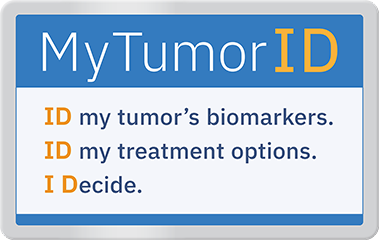
That’s why the National Brain Tumor Society launched the MyTumorID® campaign in 2024 to help educate patients, caregivers, and families about the importance of biomarker testing to receive an accurate diagnosis and make informed decisions about treatment options.
An Emergency Pediatric Brain Tumor Diagnosis
The family’s brain tumor experience began nearly a decade ago. In 2016, while living in Barcelona during her dad’s sabbatical from work as a professor, 5-year-old Marin began experiencing a variety of symptoms, including headaches, lethargy, light sensitivity, and motion sickness.
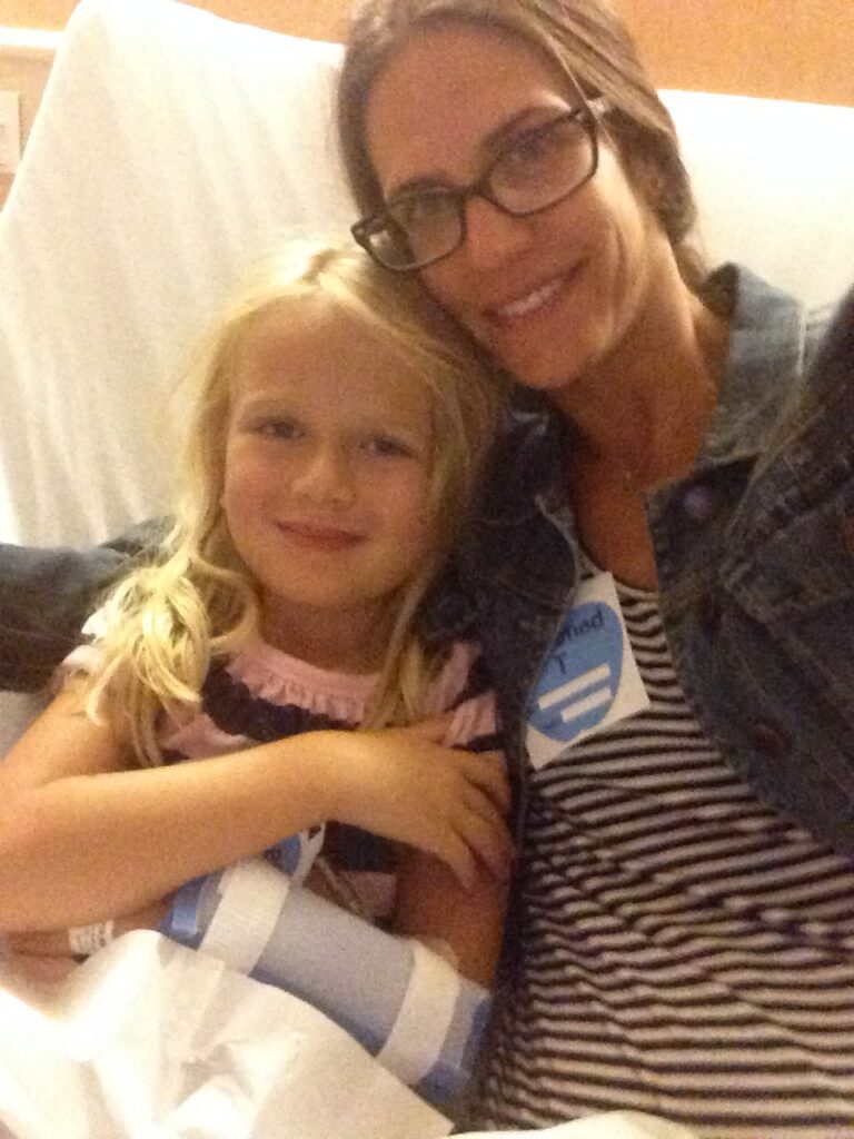
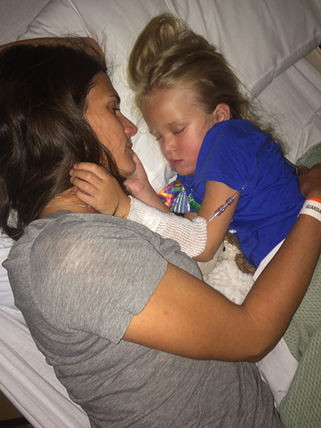
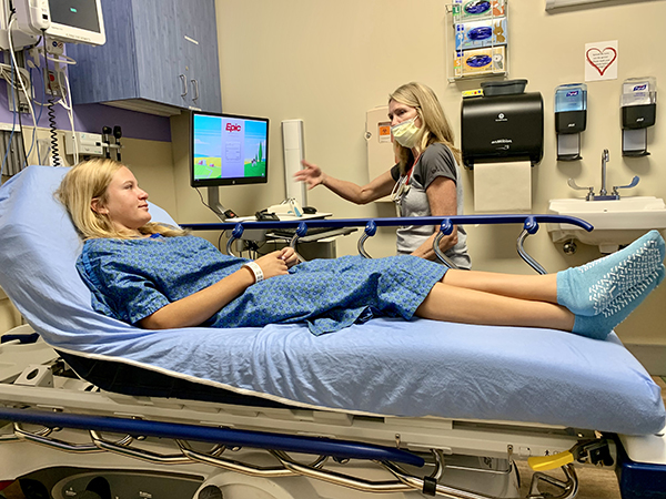
Marin & Leslie
Local pediatricians suspected a lingering virus. Shortly after returning to the United States, Marin grew dizzy and fell over while walking across the street on her way to a kindergarten meet-and-greet before the school year began.
Her mother Leslie brought Marin to a local clinic, which sent her to Children’s Hospital Colorado for a CT scan. The scan revealed a baseball-sized mass in Marin’s cerebellum, the region at the base of the brain responsible for balance, coordination, movement, and fine motor skills.
“It was like a fire hose of fear,” Marin’s father, Jeremy, recalled. “I remember my body collapsing and physically sobbing. You hear your kid has a brain tumor, and you’re worried that your kid is going to die.”
Marin underwent an emergency craniotomy to fully resect the tumor. At the time, testing was limited to screening a few basic mutations — namely the KIAA1549:BRAF fusion known to drive 65–75% of sporadic pilocytic astrocytomas — rather than the comprehensive panels used today.
Without the comprehensive testing available today, her care team provided a broad, standard pathology classification: a massively calcified low-grade glioma. They believed the tumor to be similar to juvenile pilocytic astrocytoma (JPA), which is the most common tumor found in the cerebellum.
“At the time, her oncologist told us, ‘If this tumor ever tries to grow back someday, there might be a pill that Marin could take to stop this thing in its tracks,’” Jeremy said. “I remember being blown away and thinking that it sounded like science fiction.”
The surgery was considered a success, and after a hospital recovery to treat hydrocephalus and swelling, Marin started kindergarten. For years, routine follow-up scans remained clear.
Navigating Brain Tumor Recurrence and Unanswered Questions
In 2020, four and a half years after surgery, a routine scan showed a subtle spot that doctors initially thought might be maturing scar tissue. Over the next few years, follow-up scans revealed that the tumor was slowly re-growing into the surgical cavity left behind from her first tumor resection.
Because the tumor wasn’t causing physical symptoms, her medical team closely monitored it until August 2024, when they recommended a second craniotomy. The rationale was not only to safely remove the tumor, but also to perform biomarker testing on it to figure out why it grew back in the first place.
Navigating a second surgery eight years later as parents of a now 13-year-old brought a new layer of emotional weight. Now in middle school, Marin held concerns about how surgery could impact how she looked and potential side effects that could alter her social life.
“Strangely, the second time was a lot harder because we knew all the risks,” Jeremy said. “For the first surgery, I had no role to play. This time, we had a semblance of control, which made it a little bit scarier somehow.”
Her surgery was a successful gross total resection, meaning her tumor was completely removed. Two days later, Marin returned to school.
The Biomarker Testing Turning Point
Following Marin’s 2024 surgery, her care team ordered biomarker testing on her tumor tissue. When the results came back, the family received an entirely new understanding of her tumor.
Marin did not have juvenile pilocytic astrocytoma as they had suspected in 2016. Instead, the results revealed a rare neuroepithelial tumor with an MN1:PATZ1 fusion along with a TERT promoter mutation.
While both a JPA tumor and Marin’s tumor type can look similar under a microscope, their molecular makeup is different.
A PATZ1 fusion is a rare genetic alteration occurring when part of the PATZ1 gene joins with another gene, most commonly MN1 or EWSR1 on chromosome 22.
According to an article in Genetics in Medicine Open, “PATZ1 fusion (with MN1 and EWSR1)–positive neuroepithelial tumors are uncommon but are increasingly recognized as a unique molecular subtype of neoplasms that predominantly arise in children and young adults.”
After Marin’s diagnosis, Jeremy learned that only 14 other cases with Marin’s exact MN1:PATZ1 fusion have been documented in medical literature.
“It was a bit scary at first, but it’s become more heartening to know more people are being diagnosed with this fusion and that there could be treatment that could help her if it grows back again,” Jeremy said. “Even if there aren’t medicines that can help Marin’s tumor with that fusion right now, perhaps future targeted therapies may be able to address the pathways where these processes take place.”
TERT (telomerase reverse transcriptase) is a gene involved in maintaining the ends of chromosomes, which helps regulate how cells divide. When TERT is altered (called a promoter mutation), tumor cells can continue dividing rather than stopping.
This is a prognostic marker, which is a sign that helps your care team understand how the tumor may behave over time. An alteration is often associated with more aggressive tumor behavior.
“As I understand it, a TERT promoter does not turn off the division of bad cells,” Jeremy said. “Most people don’t have a TERT promoter, so when certain cells start to grow, their body recognizes it’s bad and turns off this process. Her tumor doesn’t do that.”
In addition to some neuroepithelial tumors like Marin’s, TERTp can be found in other brain tumor types, including some oligodendrogliomas, glioblastomas, medulloblastomas, and astrocytomas.
Identifying Marin’s tumor fundamentally shifted how Jeremy and his family understood her condition and gave them a better idea of her prognosis. It explained the tumor’s growth pattern and why it returned, when JPAs typically don’t recur after a total resection. Understanding that this tumor type is more likely to return now guides long-term surveillance and care.
“Had we known that this tumor would continue to grow when we first learned of tumor regrowth, we would have gotten it out sooner,” Jeremy shared. “Had we known earlier, our emotional rollercoaster would have had fewer loops and been more straightforward. This diagnosis was incredibly helpful, not just physically for Marin, but emotionally for our family. It’s such a gift to have some control in a situation that is completely out of your control.”
Instead of relying on generalized assumptions, Marin’s medical team now monitors her health with a surveillance schedule tailored to her specific tumor characteristics. As a result, Jeremy now advocates strongly for making comprehensive biomarker testing a standard diagnostic step for every patient faced with a brain tumor diagnosis.
“It should be the standard of care in every hospital,” Jeremy said. “Biomarker testing has major impacts on the future trajectory of care for your child. I can’t say enough about how it’s changed Marin’s life and our lives as people who care the most for her.”
Blog Post Supported By
Join NBTS for a Free Biomarker Testing Webinar
When you ID your tumor through biomarker testing, you can explore treatment options with your health care team and make informed decisions about your care, including whether a clinical trial may be right for you. With MyTumorID, I Decide.
Learn more about biomarker testing when you register today for our free informative MyTumorID webinar on Oct. 2, 2026, to better understand your brain tumor biomarker testing report and what it may mean for your or your loved one’s brain tumor diagnosis.
While you wait for this new webinar, you can watch our past webinar about what biomarker testing is, how it influences treatment planning and access to clinical trials, and why it matters.