Sign our action alert today — this Glioblastoma Awareness Day, urge Congress to support GBM-specific priorities, including funding research, expanding the Glioblastoma Therapeutics Network, and co-sponsoring the BRAIN Act.
Cookie Policy
This website uses cookies that help the website function and that help us understand how you interact with it. Please read our privacy policy for more information.
The ultimate goal of scientific endeavor in the field of cancer research is to advance discoveries made in the laboratory through the translational and pre-clinical research process to become new medicines patients can take in the clinic. But as scientific discoveries and advancements occur, it is also important that results from studies be published in peer-reviewed scientific/medical journals, so that the new information can be widely disseminated to other researchers around the world to inform their work with the latest leanings.
As such, while not the ultimate measure or goal, the publication of new research papers in reputable scientific journals still serves as an important barometer of progress and successful milestones for ongoing projects to find better treatments and ultimately a cure. And today we’re happy to share with you some of the NBTS-funded research that has been published over the last few months.
The following six publications indicate the momentum being created in the brain tumor research field because of NBTS-funded studies – which we could only do with YOUR support.
Seeking Alternate Routes
Many tumors are driven by alterations to genes that cause over-activity in “signaling pathways” – the channels our cells use to communicate key messages within their walls. Thus, a key strategy to stop tumor growth in many cancers – including brain cancers like glioblastoma (GBM) – has been to use drugs to act like roadblocks within those signaling pathways to stop the increased traffic driving tumor growth. However, sometimes drugs that a researcher or clinician believe should be adequate road blocks don’t end up working. This phenomenon is known as “drug resistance.”
Researchers in recent years have begun understanding how drug resistance can be driven by other genetic mutations also occurring in the tumor cells.
Now Drs. Paul Mischel, Webster Cavenee, and Timothy Cloughsey – all members of NBTS’ Defeat GBM Research Collaborative – are part of a team that has found a new and additional way that GBM cells can resist treatment, not involving additional genetic changes.
Publishing in the newest edition of the journal Cancer Cell, the team notes that just like cars following a detour, tumors can quickly remap (or “re-wire”) their signaling pathways to find new “roads” to bypass the blockade being created by a drug.
Drug resistance was marked by the emergence of signaling networks that were previously in the background and that now were newly coordinated to drive the growth of these tumors. This was a key insight because it led to a series of testable predictions.
Attribution: Dr. Mischel
Researchers found this using new technology and techniques to visualize these alternate routes at the single cell level requiring only a small amount of tumor sample using a preclinical model of GBM, and the hopes is that by identifying these new pathways researchers will be able to cut off multiple roads at the same time. This approach could be used by clinicians to anticipate, and even overcome, drug resistance in GBM.
Title: Single-Cell Phosphoproteomics Resolves Adaptive Signaling Dynamics and Informs Targeted Combination Therapy in Glioblastoma
IDH (Isocitrate Dehydrogenase) is an enzyme that plays a key role in a cells’ citric acid cycle, which is a metabolic pathway that converts nutrients into energy as part of a cell’s metabolism. When the gene that codes for the IDH enzyme (and its protein variants) becomes altered by mutation, the enzyme lose the ability to handle its regular jobs of producing normal molecules that support cellular metabolism. Instead, the mutated enzyme adopts a new activity producing a different metabolite that accumulates and blocks the cell from functioning normally, while driving malignant progression and tumor development. IDH mutations are common in many gliomas.
In this paper from the January edition of the journal Clinical Cancer Research, Defeat GBM-funded researcher, Dr. Ingo Mellinghoff, summarizes recent progress in our understanding of the role that IDH mutations play in driving tumor development.
Title: Molecular Pathways: Isocitrate Dehydrogenase Mutations in Cancer
Epidermal growth factor receptor (EGFR) is a protein that acts essentially as a docking station on the surface of cells to receive signals from outside the cell that initiate the process for cells to grow and divide. EGFR is amplified and mutated in a large subset of glioma patients, causing cells to divide out of control, a hallmark of cancer.
The most common EGFR mutation is known as EGFRvIII. This mutation has been known for a long time, but attempts to apply treatments that counteract it have been unsuccessful to-date. Because of this, one group of researchers within NBTS’ Defeat GBM Research Collaborative has focused their efforts on better understanding the full impact the EGFRvIII mutation has on GBM cells.
In this paper from the January edition of the journal Neuro-Oncology, Defeat GBM-funded researcher Frank Furnari provides an overview of EGFR and EGFRvIII, describing therapeutic strategies to inhibit the receptor, and possible intervention routes to efficiently target this receptor and prevent the emergence of resistant mechanisms which to date have hampered a successful therapeutic outcome.
Title: Epidermal growth factor receptor targeting and challenges in glioblastoma (Frank)
Imaging Technique to Monitor Tumor Response During Treatment
As stated above, IDH mutations are common in gliomas and they cause a build up of a molecule in cells that causes the cell to cease functioning normally, leading to tumor development.
That molecule is a metabolite called “R-2-hydroxyglutarate” (R-2HG). Prior studies showed that this metabolite can be detected in animals with gliomas using imaging techniques known as proton magnetic-resonance spectroscopy (MRS). This discovery offered the possibility that researchers could use non-invasive approaches (other than repeated biopsies, which are nearly impossible to do in the brain) to monitor the levels of R-2HG in cells during treatment to see if gliomas were responding to treatment or not (a decrease in R-2HG levels in the tumor, would indicate the treatment was working).
MR image of a brain tumor
But the accuracy of this technique was not confirmed and thus the ability to use this approach in the clinic has still been in question.
Now, a team lead by Defeat GBM-funded researcher Dr. Ingo Mellinghoff has developed an MRI protocol to integrate 2HG-MRS into routine clinical glioma imaging and evaluated its performance in 89 consecutive glioma patients. The study, published recently in the journal Neuro-Oncology, demonstrates that 2HG-MRS can be linked with routine MR imaging to provide quantitative measurements of 2HG in glioma and may be useful as an imaging biomarker to monitor the abundance of IDH-mutant tumor cells noninvasively during glioma therapy and disease monitoring, especially for larger tumors. Additionally improving imaging technology in the future can help make this approach even more accurate, particularly for smaller tumors.
Title: Integration of 2-Hydroxyglutarate-Proton Magnetic Resonance Spectroscopy into Clinical Practice for Disease Monitoring in Isocitrate Dehydrogenase-Mutant glioma
BRAF is a gene that makes a protein called B-Raf. The B-Raf protein is involved in sending signals inside cells, which are involved in directing cell growth. A faulty BRAF gene has been found in over 80% of sporadic pilocytic astrocytomas.
In these tumors the BRAF gene is erroneously duplicated, which creates a “fusion” version of the B-Raf protein. The fusion version of the B-Raf protein, known as BRAF-KD, is unable to maintain its normal function, and the signals that it sends telling cells to grow stays on indefinitely and also activates another signaling pathway know as MAPK.
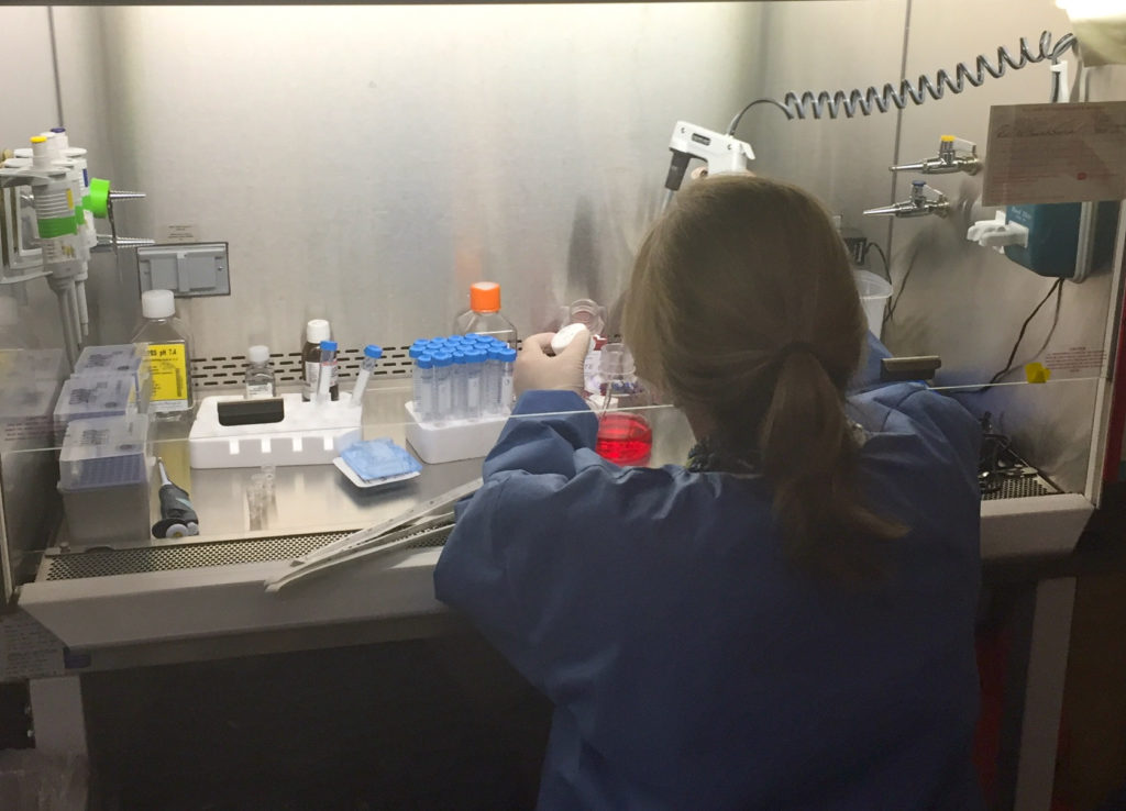
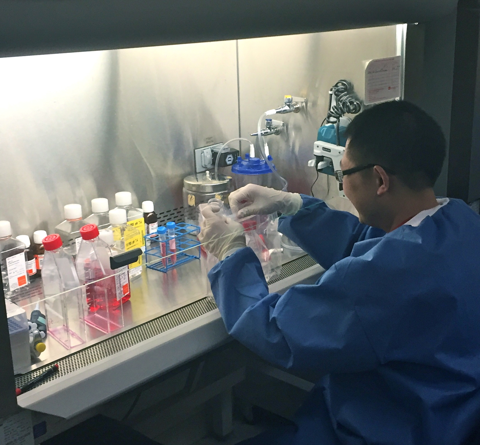
Inside the Neuro-Oncology lab at the NIH/NCI
NBTS-funded researcher Dr. Sheri Holmen and a team of other scientists set out to determine if BRAF-KD alone can cause gliomas, or if it needs to act in cooperation with another molecular change for a tumor to form.
The team’s findings, published in the January issue of the journal Genes & Cancer, found that for glioma formation in a mouse model, the BRAF-KD needs to be accompanied by a deletion (causing loss of function) within a spot on a chromosome (chromosome 9) known as INK4A/ARF. Additionally, they found that the cells with BRAF-KD and INK4A/ARF loss that turn into tumors can often be determined by the level of MAPK signaling in the cell.
This finding gives us a better understanding on how some pilocytic astrocytomas may develop and could eventually lead to new treatment strategies.
Title: The BRAF Kinase Domain Promotes the Development of Gliomas In Vivo
Drs. Mario Suva and David Louis, both of Massachusetts General Hospital (MGH), together received one of the first two research grants NBTS gave out as part of our Oligodendroglioma Research Fund. Their grant project was aimed at identifying ways that oligodendroglioma and other low-grade gliomas develop, and provide information on potential targets and biomarkers for future treatment development and disease monitoring.
Collaborating with other researchers from the Broad Institute and MGH, Dr. Suva has contributed to a study that proposes a new mechanism for glioma development and growth. The study was published in the world-renowned journal Nature in early January. The research uncovered changes in the instructions for how the genome folds up on itself in IDH-mutant gliomas. The changes affect parts of the genome, called insulators, which physically prevent genes in one region from interacting with the control switches and genes that lie in neighboring regions. Disruption of a specific boundary bordering a growth factor gene known as PDGFRA activated this gene to promote glioma formation and growth. The study also suggests that the PDGFRA gene is unlikely to be the only gene affected by altered genome folding and changes in “insulation”.
This is a totally new mechanism for causing cancer, and we think it will hold true not just in brain tumors, but in other forms of cancer. It is well established that cancer-causing genes can be abnormally activated by changes in their DNA sequence. But in this case, we find that a cancer-causing gene is switched on by a change in how the genome folds.
Attribution: Senior author Bradley Bernstein, institute member at the Broad Institute and pathology professor at Massachusetts General Hospital
Title: Insulator Dysfunction and Oncogene Activation in IDH Mutant Gliomas